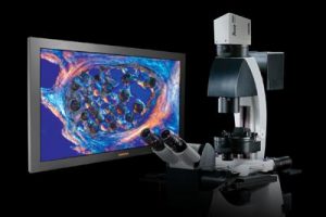
maging system in order to improve the visualisation of patient data for doctors, surgeons, pathologists and research scientists.

The Imaris 3D system incorporates features that allow time-based visualisation of images, the creation of 3D or multi-channel images, or the production of animation movies. One feature, for instance, can be used to monitor temporal changes in biological systems. The system also seeks out filaments such as neurons, microtubules or blood vessels and retains and calculates topological information for the user.
The Imaris system can also be used in conjunction with a stereomicroscopes or endoscopes either to get a real-time and deeper view of patient tissue or as a training tool for future surgeons.
